

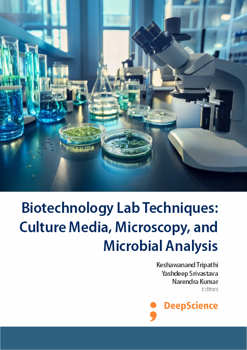
Paraffin block preparation and histological staining: Techniques for tissue structure analysis
Synopsis
Fixation is vital for preserving tissue integrity for histological examination, aiming to maintain tissues in a lifelike state. Immediate fixation, post-surgery or autopsy, is crucial to prevent autolysis. Tissues are prepared for transformation into thin microscopic slices following fixation. One important method is to embed tissues in paraffin that matches their density. Using a microtome, paraffin-embedded tissues are cut into micrometer-thin slices, with paraffin serving as support. However, in order for water- and alcohol-soluble dyes to penetrate, paraffin must be eliminated during staining. The Hematoxylin and Eosin stain, commonly used in histology, gives distinct coloration to cellular components. Hematoxylin, which binds to nucleic acids, stains cell nuclei bluish-purple. Eosin, binding to cytoplasmic elements, imparts a pink hue to the background. Hematoxylin requires a mordant to enhance staining affinity. Mordants, high-molecular-weight metal salts, form strong bonds with tissues and dyes, ensuring effective staining.
Downloads
Published
13 April 2025
Copyright (c) 2025 Keshawanand Tripathi; Yashdeep Srivastava, Narendra Kumar
How to Cite
Tripathi, K. ., Srivastava, Y. ., & Kumar, N. . (2025). Paraffin block preparation and histological staining: Techniques for tissue structure analysis. In K. . Tripathi, Y. . Srivastava, & N. . Kumar (Eds.), Biotechnology Lab Techniques: Culture Media, Microscopy, and Microbial Analysis (pp. 69-74). Deep Science Publishing. https://doi.org/10.70593/978-93-49307-52-0_11
Download Citation


 WhatsApp +91 7977171947

